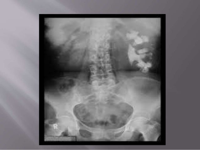
If left untreated, staghorn calculi result in chronic infection and eventually may progress to xanthogranulomatous pyelonephritis 5. Staghorn calculi need to be treated surgically, usually PCNL (percutaneous nephrolithotomy) +/- ESWL (extracorporeal shockwave lithotripsy) and the entire stone removed, including small fragments, as otherwise, these residual fragments act as a reservoir for infection and recurrent stone formation.
Staghorn calculus meme windows#
When viewed on bone windows they have a laminated appearance, due to alternating bands of magnesium ammonium phosphate and calcium phosphate 5.

Staghorn calculi are radiopaque and conform to the renal pelvis and calyces, which are often to some degree dilated. The collecting system is filled with a densely calcified mass, producing marked posterior acoustic shadowing. The vast majority of staghorn calculi are radiopaque and appear as branching calcific densities overlying the renal outline and may mimic an excretory phase intravenous pyelogram. They refer to struvite calculi involving the renal pelvis and extending into at least two calyces 7. However, a staghorn calculus is generally consid-ered a branching renal stone that occupies multiple portions of the renal collecting system. Staghorn calculi, also sometimes called coral calculi, are renal calculi that obtain their characteristic shape by forming a cast of the renal pelvis and calyces, thus resembling the horns of a stag. tial versus a complete staghorn calculus, such as vol-ume criteria or number of calyces occupied. Uric acid and cystine are the underlying components of a minority of these calculi 5. Citation, DOI, disclosures and article data. Struvite accounts for approximately 70% of the composition of these calculi and is usually mixed with calcium phosphate thus rendering them radiopaque on both plain films and CT. Urease hydrolyzes urea to ammonium with an increase in the urinary pH 3-5. Proteus, Klebsiella, Pseudomonas and Enterobacter). Staghorn calculi are composed of struvite (chemically this is magnesium ammonium phosphate or MAP) and are usually seen in the setting of recurrent urinary tract infection with urease-producing bacteria (e.g. The majority of staghorn calculi are symptomatic, presenting with fever, hematuria, flank pain and potentially septicemia and abscess formation. doi: 10.1111/j.Staghorn calculi are the result of recurrent infection and are thus more commonly encountered in women 6, those with renal tract anomalies, reflux, spinal cord injuries, neurogenic bladder or ileal ureteral diversion. Fibrosis and evidence for epithelial-mesenchymal transition in the kidneys of patients with staghorn calculi. 1993 22(4):513–519.īoonla C, Krieglstein K, Bovornpadungkitti S, Strutz F, Spittau B, Predanon C, Tosukhowong P. Renal stone disease in autosomal dominant polycystic kidney disease.

Torres VE, Wilson DM, Hattery RR, Segura JW. Lithiasis in cystic kidney disease and malformations of the urinary tract. Evaluation of nephrolithiasis in autosomal dominant polycystic kidney disease patients. Nishiura JL, Neves RF, Eloi SR, Cintra SM, Ajzen SA, Heilberg IP. Staghorn calculi themselves do not typically produce symptoms unless the stone results in urinary tract obstruction. This is a situation in which a patient could become septic very quickly if left untreated. Nephrolithiasis in autosomal dominant polycystic kidney disease. Kidney stones in adults: Diagnosis and acute management of suspected nephrolithiasis. As staghorn calculi are associated with kidney fibrosis and high long-term renal deterioration rate, prompt control of urinary tract infection in polycystic kidney disease patient will be beneficial in preventing staghorn calculus formation. UTI is an important complication for polycystic kidney disease and will facilitate the formation of staghorn calculi.

CT scan revealed the existence of a complete staghorn calculus in her right kidney, while there was no kidney stone 3 years before, and the urinary stone component analysis showed the composition of calculus was magnesium ammonium phosphate. This 37-year-old autosomal dominant polycystic kidney disease female with positive family history was admitted in this hospital for repeatedly upper urinary tract infection for 3 years.

We report a case of complete staghorm calculus in a polycystic kidney disease patient induced by repeatedly urinary tract infections. For general population, recent data showed metabolic factors were the dominant causes for staghorn calculus, but for polycystic kidney disease patients, the cause for staghorn calculus remained elusive. However, complete staghorn calculus is rare in polycystic kidney disease and predicts a gloomy prognosis of kidney. Kidney stones in patients with autosomal dominant polycystic kidney disease are common, regarded as the consequence of the combination of anatomic abnormality and metabolic risk factors.


 0 kommentar(er)
